Interactive veterinary anatomy
Atlas atlas.pisoro.com
Two independent builds are hosted here, each a self-contained static viewer with its own geometry pipeline, sources and licence record. Neither replaces the other: one shows a whole animal, the other shows what each structure was actually derived from.
Builds
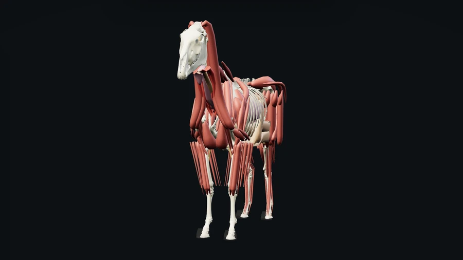
Specimen study
Whole-body atlas
/study/
Two species side by side, each built from one specimen: articulated skeleton, muscles placed on their own model's attachment paths, and modelled viscera fitted inside the real body cavity. Layer, region and side filters, Latin terminology, ranked clinical search.
- Species
- 2
- Structures
- 510
- Muscles
- 302
- Organs
- 78
- Joints
- 31
Horse SHADOWFAX model, van Bijlert et al. 2024 (MIT) · published lines of action and
dissection masses.
Dog Stark et al. 2016 canine model, standing pose solved from the model's own gait
trials.
Viscera modelled, not scanned.
Open the whole-body atlas
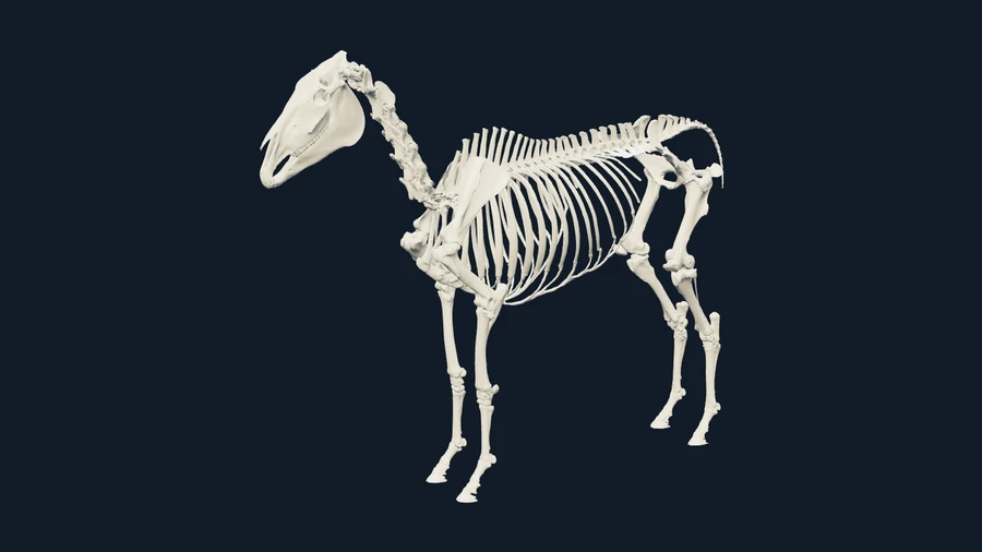
Source imaging
Scans & evidence
/imaging/
The scanned skeleton at source resolution with muscles and organs registered onto it, beside five independent published datasets kept deliberately unmerged — with the native scan sections and their original segmentation overlays viewable next to the surfaces. Every label carries its provenance, review state and the reference it still needs before it can be trusted.
- Datasets
- 6
- Structures
- 357
- Scanned
- 170labels
- Pending
- 365refs
Skeleton SHADOWFAX full skeleton (25 surfaces) · EquiSim distal forelimb (10).
Imaging TEBTA head CT (8) · TEBTA brain MRI (115) · incisor CT, subject k1 (12).
Soft tissue 187 modelled structures registered to the skeleton by a measured translation,
plus 14 externally hosted dissection specimens.
Open the imaging atlas
Which one to open
For teaching a whole animal — where a muscle runs, what a region contains, how two species differ — open the whole-body atlas. To work from the scan a structure was actually segmented from, open the imaging atlas.
What is shared
Both use the same published horse skeleton, so the muscles and organs built here are carried into the imaging atlas by a measured translation with zero residual, not an estimated fit. The scan datasets stay unregistered to each other and to it — that alignment nobody has verified.
Status
Research builds, not clinically validated, and neither is a complete animal. Sources and derivatives keep their own licences and attribution; each build links its machine-readable provenance record.